Publications

Actively Multiplexed μECoG Brain Implant System With Incremental- ΔΣ ADCs Employing Bulk-DACs

Fundamental neuroscience research and high-performance neuro-prostheses require large-scale brain interfaces with ever-greater spatial resolution across a large cortex coverage, which cannot be achieved with current passive (micro) electrocorticography (ECoG) technologies. In this article, we present an active micro-electrocorticography (μECoG) implant system that circumvents these challenges while achieving significantly lower noise compared to other existing active μECoG arrays. The proposed brain implant system is composed of a flexible, actively multiplexed 256-electrode μECoG array and an incremental-ΔΣ readout integrated circuit (ROIC). The 1 cm x 1 cm μECoG array was fabricated in a 3-μm IGZO thin-film transistor (TFT) technology on a 15-μm flexible foil and coupled to a 1.25 mm x 1.25 mm CMOS ROIC fabricated in a 22-nm fully depleted silicon on insulator (FDSOI) process. Due to the 256:16 time-division multiplexing achieved in the electrode array, only 16 multiplexed channels are required in the ROIC to acquire signals from the 256 electrodes simultaneously. By combining TFT multiplexing with newly proposed bulk-DAC (BDAC) feedback in the readout channel, we can integrate and address 4x more electrodes than other passive arrays, achieve >10x less noise than existing active arrays, and obtain >2x effective channel area reduction in the ROIC while maintaining comparable electrical performance over current state-of-the-art (μ)ECoG readouts.

Implantation of Neuropixels probes for chronic recording of neuronal activity in freely behaving mice and rats

How dynamic activity in neural circuits gives rise to behaviour is a major area of interest in neuroscience. A key experimental approach for addressing this question involves measuring extracellular neuronal activity in awake, behaving animals. Recently developed Neuropixels probes have provided a step-change in recording neural activity in large tissue volumes with high spatiotemporal resolution. This protocol describes the chronic implantation of Neuropixels probes in mice and rats using compact and reusable 3D-printed fixtures. The fixtures facilitate stable chronic in-vivo recordings in freely behaving rats and mice. They consist of two parts: a covered main body and a skull connector. Single, dual, and movable probe fixture variants are available. After completing an experiment, probes are safely recovered for reimplantation by a dedicated retrieval mechanism. Fixture assembly and surgical implantation typically take 4-5 h, probe retrieval takes about half an hour, followed by a 12 h incubation in probe cleaning agent. The duration of data acquisition depends on the type of behavioral experiment. Since our protocol enables stable, chronic recordings over weeks, it enableslongitudinal large-scale single-unit data to be routinely obtained in a cost-efficient manner, which will facilitate many studies in systems neuroscience.

Neuropixels 2.0: A miniaturized high-density probe for stable, long-term brain recordings

To study the dynamics of neural processing across timescales, we require the ability to follow the spiking of thousands of individually separable neurons over weeks and months, during unrestrained behavior. To address this need, we introduce the Neuropixels 2.0 probe together with novel analysis algorithms. The new probe has over 5,000 sites and is miniaturized such that two probes plus a headstage, recording 768 sites at once, weigh just over 1 g, suitable for implanting chronically in small mammals. Recordings with high quality signals persisting for at least two months were reliably obtained in two species and six different labs. Improved site density and arrangement combined with new data processing methods enable automatic post-hoc stabilization of data despite brain movements during behavior and across days, allowing recording from the same neurons in the mouse visual cortex for over 2 months. Additionally, an optional configuration allows for recording from multiple sites per available channel, with a penalty to signal-to-noise ratio. These probes and algorithms enable stable recordings from >10,000 sites during free behavior in small animals such as mice.Competing Interest StatementThe authors have declared no competing interest.

Proximal and distal modulation of neural activity by spatially confined optogenetic activation with an integrated high-density optoelectrode

Optogenetic manipulations are widely used for investigating the contribution of genetically identified cell types to behavior. Simultaneous electrophysiological recordings are less common, although they are critical for characterizing the specific impact of optogenetic manipulations on neural circuits in vivo. This is at least in part because combining photostimulation with large-scale electrophysiological recordings remains technically challenging, which also poses a limitation for performing extracellular identification experiments. Currently available interfaces that guide light of the appropriate wavelength into the brain combined with an electrophysiological modality suffer from various drawbacks such as a bulky size, low spatial resolution, heat dissipation, or photovoltaic artifacts. To address these challenges, we have designed and fabricated an integrated ultrathin neural interface with 12 optical outputs and 24 electrodes. We used the device to measure the effect of localized stimulation in the anterior olfactory cortex, a paleocortical structure involved in olfactory processing. Our experiments in adult mice demonstrate that because of its small dimensions, our novel tool causes far less tissue damage than commercially available devices. Moreover, optical stimulation and recording can be performed simultaneously, with no measurable electrical artifact during optical stimulation. Importantly, optical stimulation can be confined to small volumes with approximately single-cortical layer thickness. Finally, we find that even highly localized optical stimulation causes inhibition at more distant sites.NEW & NOTEWORTHY In this study, we establish a novel tool for simultaneous extracellular recording and optogenetic photostimulation. Because the device is built using established microchip technology, it can be fabricated with high reproducibility and reliability. We further show that even very localized stimulation affects neural firing far beyond the stimulation site. This demonstrates the difficulty in predicting circuit-level effects of optogenetic manipulations and highlights the importance of closely monitoring neural activity in optogenetic experiments.

IR thermography-based monitoring of respiration phase without image segmentation

Background Respiratory rate is an essential parameter in biomedical research and clinical applications. Most respiration measurement techniques in preclinical animal models require surgical implantation of sensors. Current clinical measurement modalities typically involve attachment of sensors to the patient, causing discomfort. We have previously developed a non-contact approach to measuring respiration phase in head-restrained rodents using infrared (IR) thermography. While the non-invasive nature of IR thermography offers many advantages, it also bears the complexity of extracting respiration signals from videos. Previously reported algorithms involve image segmentation to identify the nose in IR videos and extract breathing-relevant pixels which is particularly challenging if the videos have low contrast or suffer from suboptimal focusing. New method To address this challenge, we developed a novel algorithm, which extracts respiration signals based on pixel time series, removing the need for nose-tracking and image segmentation. Results & comparison with existing methods We validated the algorithm by performing respiration measurements in head-restrained mice and in humans with IR thermography in parallel with established standard techniques. We find the algorithm reliably detects inhalation onsets with high temporal precision. Conclusions The new algorithm facilitates the application of IR thermography for measuring respiration in biomedical research and in clinical settings.

Spontaneous Rapid Odor Source Localization Behavior Requires Interhemispheric Communication

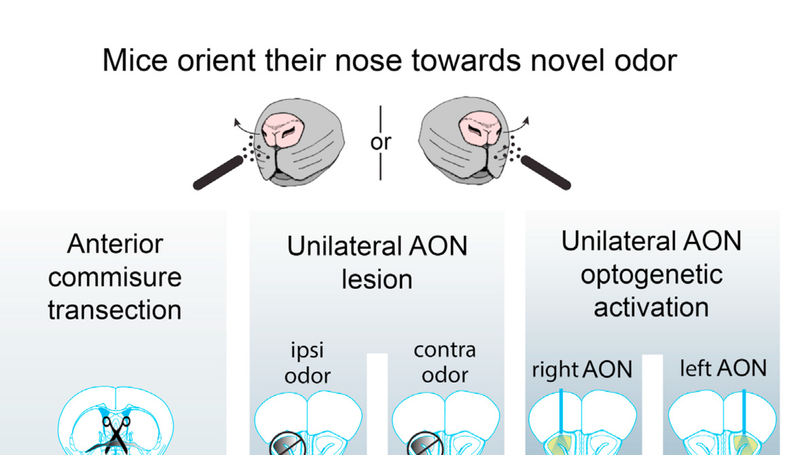
Summary Navigation, finding food sources, and avoiding danger critically depend on the identification and spatial localization of airborne chemicals. When monitoring the olfactory environment, rodents spontaneously engage in active olfactory sampling behavior, also referred to as exploratory sniffing [1]. Exploratory sniffing is characterized by stereotypical high-frequency respiration, which is also reliably evoked by novel odorant stimuli [2, 3]. To study novelty-induced exploratory sniffing, we developed a novel, non-contact method for measuring respiration by infrared (IR) thermography in a behavioral paradigm in which novel and familiar stimuli are presented to head-restrained mice. We validated the method by simultaneously performing nasal pressure measurements, a commonly used invasive approach [2, 4], and confirmed highly reliable detection of inhalation onsets. We further discovered that mice actively orient their nostrils toward novel, previously unexperienced, smells. In line with the remarkable speed of olfactory processing reported previously [3, 5, 6], we find that mice initiate their response already within the first sniff after odor onset. Moreover, transecting the anterior commissure (AC) disrupted orienting, indicating that the orienting response requires interhemispheric transfer of information. This suggests that mice compare odorant information obtained from the two bilaterally symmetric nostrils to locate the source of the novel odorant. We further demonstrate that asymmetric activation of the anterior olfactory nucleus (AON) is both necessary and sufficient for eliciting orienting responses. These findings support the view that the AON plays an important role in the internostril difference comparison underlying rapid odor source localization.